Multiphoton microscopy
This imaging technique is used to monitor in a label-free manner the non-linear optical response (two-photon excited fluorescence and second harmonic generation) of biomolecular systems like collagen, microtubules, myofibrils, sarcomeres in very varied biological structures such as cancerous cells, cardiomyocytes, stem cells, excised spinal cord, muscle or cardiac tissues.
SHG signal analysis and processing algorithms developed in our laboratory are used to compute and quantify density, length, tortuosity and orientation of myofibrils, sarcomeres and collagen fibers.
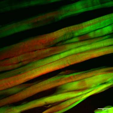
Sarcomeres organization in a mice tibia muscle (green – SHG signal, red – two-photon excited fluorescence of the tissue). Image recorded in multiphoton microscopy.
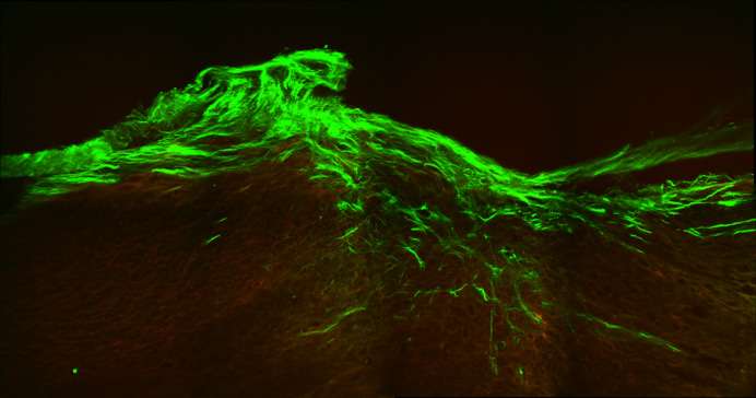
Collagen fibers growth in lesioned spinal cord tissue in a murine model (green – SHG signal, red – two-photon excited fluorescence of the tissue and labeled astrocytes). Image recorded in multiphoton microscopy.